Anatomy & Physiology
Bauplan
Placida dendritica differs from the characteristic molluscan bauplan (body plan) in a number of ways. Firstly, P.dendritica is detorted, meaning that the anus and posterior of the animal is no longer above the head, as in most other gastropod molluscs. Furthermore, P. dendritica, as a sacoglossan sea slug, has secondarily lost its protective outer shell, a characteristic of most molluscan taxa.
P. dendritica is a coelomate organism (ie. contains a coelom, or internal compartment, which is separate from the gut) with a haemocoel cavity through which blood circulates. These haemocoelic spaces surround longitudinal and transverse muscles (Marzo et al. 1993). P. dendritica has secondarily evolved bilateral symmetry (Ruppert et al. 2004). Externally, it exhibits this symmetry almost perfectly, however, the internal symmetry has a slight skew to the right. This is due to the detorsion process (Ruppert et al. 2004). As a result, some organs, such as the reproductive system, are located more on the right side of the body. Aspects of P. dendritica reproductive, digestive, respiratory and nervous systems are detailed in the sections below.
The general anatomical layout of P. dendritica is presented in Figure 1and Photo (a) (see below). The buccal mass, oesophagus, stomach and anus form the digestive system. The heart is located just behind the stomach. For more information on the digestive system, see section below: Digestive System. The buccal mass includes the mouth, pharynx and radula sac (an outpocket underneath the pharynx which contains the radula). The radula (see section below: Radula) is a row of chitinous teeth used in feeding. The radula sac is a highly mineralized region of the body, and as such, shows up on H&E stain (haematoxylin and eosin stain) slides as bright orange. A cross section taken through the body of a P. dendritica specimen from Heron Island, Australia, is believed to have captured the radula sac, sitting below an apparent digestive gland (see Photo (a)). Many features in this stain are yet to be identified, however the digestive tract and muscular foot are recognisable. Cerata can be seen around the dorsal surface of the animal.
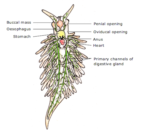
Figure 1: Dorsal view of Placida dendritica showing the positions of various anatomical features (labelled): buccal mass, which includes the radula and pharynx (orange); oesophagus; stomach (yellow); heart (red); anus, penial and oviducal openings; and the two primary channels of the digestive system. Drawing by Alison Carlisle, adapted from illustration by I. Smith, and based on anatomical descriptions by Alder &Hancock (1843), and Baba (1986).
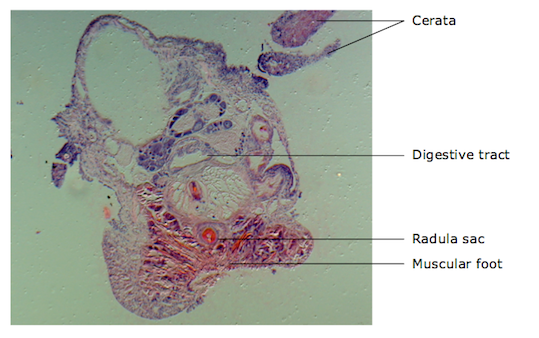
Photo (a): Cross section through the body of Placida dendritica, showing various anatomical features. The cerata, digestive tract, (tentatively identified) radula sac, and muscular foot are labelled. Photo taken by Alison Carlisle at the University of Queensland, St Lucia, Australia.
Digestive System
The digestive system of Placida dendritica is concentrated at the anterior end of the organism. The mouth is located at the most anterior point, and is connected to the pharynx and the radula sac, which contains the radula (see section below: Radula). The pharynx is connected to the mouth via a short oral tube, and from the pharynx a straight, narrow oesophagus leads to the stomach (see Figure 1) (Glascoigne, 1979). From the stomach, two short digestive channels extend into the head and rhinophores, whilst two long, primary digestive channels extend from the stomach all the way to the tail of the animal. From these channels spread a vast network of tributaries into the cerata. The amount of branching of tributaries varies between individuals. The albumen gland (a yolky gland of the reproductive system) also extends into the cerata, following the digestive tracts. This can be seen in Photo (b) (a longitudinal section of a ceras), along with nuclei and the epithelial cells (skin) which line the surface of the ceras (plural: cerata). The digestive system is green in colouration, as P. dendritica has the capacity to retain the chloroplasts from the algae it ingests (see Life History and Behaviour tab: Feeding). The anal opening is on the dorsal surface behind the head and in front of the heart (Alder & Hancock, 1843).
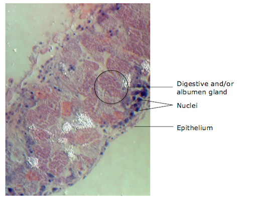
Photo (b): Close-up of a longitudinal section of a Placida dendritica ceras, showing cells of the digestive and/or albumen gland, nuclei (purple), and epithelial cells (labelled). Photo taken by Alison Carlisle at the University of Queensland, St Lucia, Australia.
Reproduction
Reproductive Organs
Placida dendritica is hermaphroditic (see Life History and Behaviour tab: Reproduction). The penial opening is located on the right side of the head, just below, and a little in front of the eye (see Figure 1). The penis extends through the penial opening during mating, and its tip is armed with a style (a pointed, hollow extension of the vas deferens) (see Figure 2). The style of P. dendritica is very small, about 40-60μm in length, and is kept in a hollow at the tip of the penis when not in use (Gascoigne, 1979). The style is used to transfer sperm to the receiving individual by hypodermic injection. In general, injection is through the skin into either the receptive organ (the bursa copulatrix) or anywhere else on the dorsal surface of the receiver’s body (Valerie et al. 2007). However, the bursa copulatrix of P. dendritica is located close to the skin surface, and thus it has been thought that this is where the sperm is received during copulation (Gascoigne, 1974).
The oviducal opening, through which the eggs are passed from the ovaries, is located a short distance behind the penial opening, just in front of the most anterior cerata on the right hand side (see Figure 1) (Jensen, 1996). The seminal receptacle, a vesicle that stores sperm, is located close to the oviducal opening and the bursa copulatrix (Jensen, 1996).
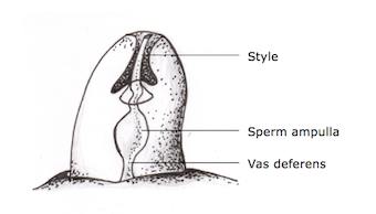
Figure 2: A generalized penetrant penial style with labels showing style, vas deferens and sperm ampulla (storage vesicle). Drawing by Alison Carlisle, adapted from illustration by Gascoigne (1973).
Eggs and Larvae
Eggs are laid with a thick gelatinous covering that is transparent, in coiled clusters on algal hosts (Alder & Hancock, 1845). Individual eggs are very small, averaging at 72.0 ±5.1μm (Clark, 1975). Larvae are planktotrophic, are transparent and have ciliated oral lobes (see Figure 3 below).
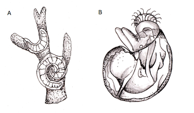
Figure 3: The egg mass (A) and larvae (B) of Placida dendritica. Drawings by Alison Carlisle, adapted from illustrations by Alder & Hancock (1845).
Radula
The radula, sometimes referred to as the ‘tongue’, is a longitudinal ribbon containing rows of chitinous teeth, which is used in feeding (see Figure 4). This structure is unique to molluscs. The radula teeth of P. dendritica are blade-shaped (as opposed to chisel-shaped) and are used for puncturing and cutting algal cell walls (see Figure 5). Interestingly, the size and number of radula teeth differs between P. dendritica individuals based on the algal species from which each individual feeds. Individuals that feed on Bryopsis species tend to have fewer, larger teeth than those that consume Codium species (Bleakney, 1989).
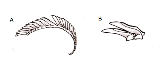
Figure 4: Generalised radula of P.dendritica (A), and close-up of two interlocking, blade-shaped radula teeth (B). Drawing by Alison Carlisle based on illustrations by Baba (1986).
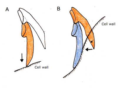
Figure 5: The piercing and cutting action of a Placida dendritica radula. (A) the cell wall is pierced by the tip of the tooth (in orange). (B) the tooth (in orange) then cuts the cell wall from the inside, as it comes into contact with the tooth in front of it (in blue). Arrows indicate direction of tooth movement and force. Drawing by Alison Carlisle, adapted from illustration by Jensen (1993).
Respiratory system
Placida dendritica, as a sacoglossan sea slug, exchanges gases with its environment through its dorsal surface (Clark, 1994). This is due to its many cerata, which considerably increase the surface area for respiration as well as for digestion (see Physical Description tab for a diagram of the dorsal surface).
Nervous System
Placida dendritica has a central nervous system that is more or less bilaterally symmetrical. The cerebral ganglia control messages from the eyes and rhinophores, and are connected to the pedal ganglia, which control the muscular foot. Together, these ganglia form a nerve ring around the oesophagus, just behind the buccal mass (see Figure 6) (Ruppert et al. 2004; Jensen, 1996). The large nerves extending from the cerebral ganglia to the well-developed eyes and chemosensitive rhinophores allow these animals to sense their surrounding environment through sight and smell.
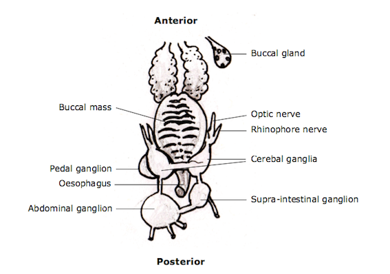
Figure 6: Dorsal view of the Central Nervous System of Placida dendritica. Various major ganglia, organs and nerves are labeled. Drawing by Alison Carlisle, adapted from illustration by Baba (1986).
Muscular Foot
The foot is a flat and highly muscular organ used in locomotion. Movement occurs through the contraction of muscles, which produces waves running from the anterior to the posterior end of the sole (middle portion) of the foot. The foot also contains many glands that secrete mucus, which aids in locomotion. Muscle fibres and (potential) glands can be seen in the H&E stain below (Photo (c)). Muscle fibres are long and extend away from the base of the foot. Glands are identified by aggregations of cells with nuclei (purple) around a channel.
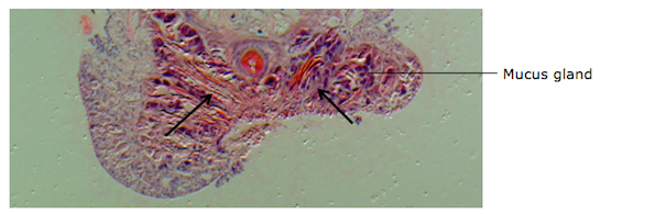
Photo (c): Close up of muscular foot from a cross-section of Placida dendritica. Arrows point to linear muscle fibre arrangements, and a potential mucus gland is labelled. Photo taken by Alison Carlisle at the University of Queensland, St Lucia, Australia.
|